ITEM SPECIFICS
-
Brand
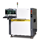
Model PS-230, PS-250Unbranded
-
origin
Republic of Korea
-
Size(Capacity)
PRODUCT DESCRIPTION
|
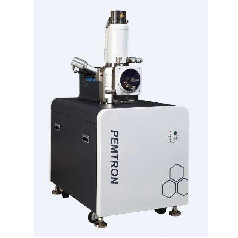
Introduction
* Electron Optic System
* Display System
|
||||||||||||||||||||||||||||||||||||||||||||||||||||||||
R&D CERTIFICATE
PAYMENTS DETAILS
- Telegraphic Transfer : T/T
- Name : Yoo YoungWoong
SHIPPING
- 219 Gasan digital 1-ro, Geumcheon-gu, Seoul (08501)
The person in charge
YoungWoong YooAddress
219 Gasan digital 1-ro, Geumcheon-gu, Seoul (08501)
-
- Business Type :
- Manufacturer
-
- Main Product :
- ATHENA
-
- Established :
- 2002-01-15
-
- Total Annual Revenue :
- More than 10 billion (KRW)
-
- Total Employees :
- 101~500 people
Please suggest a variety of your ideas such as design, impact, enhancements, etc
Please enter the text on the left image to prevent automatic input.
0 / 4000
CUSTOMER REVIEWS (0)

TRADE EXPERIENCE
-
- Total revenue
- More than 10 billion (KRW)
-
- Total export revenue (previous year in USD)
- 28,024
-
- Number of foreign trade employees
- 101~500 people
COMPARISON TO SIMILAR ITEMS more
- No Items
- supplier level
-
 SILVER
- PEMTRON Corporation Seller's Store
- Seller's Store url
- Response Level
★ ★ ★ ★ ★

- Supplier Level
★ ★ ★ ★ ★
- Transaction Level
★ ★ ★ ★ ★